- Function of synovial joints, for example, the hip or the knee, depends on the unique mechanical properties of the articular cartilage that forms their bearing surfaces.
- It distributes loads, thereby minimizing stresses on subchondral bone.
- When loaded, it deforms and when unloaded, it regains it original shape.
- It provides a surface with almost unequalled gliding properties and has remarkable durability.
- Although only a few millimeters thick, articular cartilage has an elaborate internal organization.
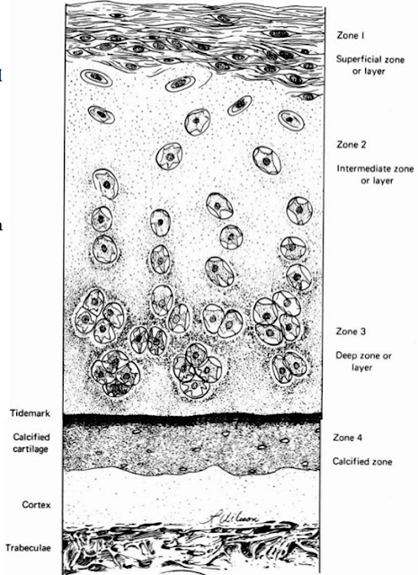
- This organization can be described by dividing articular cartilage into four successive zones beginning at the joint surface:
- The Superficial Or Gliding Zone
- The Intermediate, Middle Or Transitional Zone
- The Deep Or Radial Zone
- The Calcified Cartilage Zone
- Within zones, differences in matrix composition and organization distinguish 3 regions:
- The Pericellular Region
- The Territorial Region
- The Interterritorial Region.
Cartilage Zones
Cartilage zones differ in matrix composition, water concentration, collagen fibril orientation and cell alignment, and morphology.
1) Superficial Zone.
- The superficial or gliding zone, the smallest cartilage zone, forms the joint surface.
- A thin cell-free layer of matrix, containing primarily fine fibrils, lies directly next to the synovial cavity.
- Deep to this layer, elongated flattened chondrocytes surrounded by a larger volume of matrices per cell align their major axes parallel to the articular surface.
- The collagen fibrils of this zone lie roughly parallel to the articular surface.
2) Middle Zone
- The middle or transition zone has several times the volume of the superficial zone.
- Its more spherical cells contain greater vol of ER, Golgi membranes, mitochondria, and glycogen.
- The larger interterritorial matrix collagen fibrils of the transitional zone have a more random orientation than those of the gliding zone.
3) Deep Zone
- The cells of the deep or radial zone resemble the spherical cells of the transitional zone but tend to align themselves in columns.
- This zone has the largest collagen fibrils, highest proteoglycan content, & the lowest water content.
4) Calcified Cartilage Zone
- The thin calcified cartilage zone separates the hyaline cartilage from the stiffer subchondral bone.
- Collagen fibrils penetrate from the deep zone of cartilage directly through calcified cartilage into bone, thereby anchoring articular cartilage to subchondral bone.
Matrix Regions
Matrix regions differ in their proximity to chondrocytes, collagen content, collagen fibril diameter, collagen fibril orientation, and proteoglycan and noncollagenous protein content and organization.
1) Pericellular Matrix.
- The smallest matrix compartment, the pericellular matrix, consists of a thin layer of matrix containing little or no fibrillar collagen.
- It appears to attach directly to the chondrocyte cell membranes and probably contains noncollagenous proteins that help chondrocytes bind themselves to the matrix.
2) Territorial Matrix.
- An envelope of territorial matrix surrounds the pericellular matrix and sometimes pairs or clusters of chondrocytes and their pericellular matrices.
- Thin collagen fibrils in the territorial matrix near the cells appear to bind to the pericellular matrix.
- At a distance from the cell they spread and intersect at various angles, forming a basket like structure around the cells.
3) Interterritorial Matrix.
- The interterritorial matrix, the largest matrix compartment, has collagen fibrils of greater diameter than the territorial matrix.
- The organization and orientation of these interterritorial collagen matrix fibrils change as they pass from the articular surface to the deep region of the cartilage.
- In the most superficial zone they lie primarily parallel to the joint surface, in the transition zone they assume a more random orientation, and in the deep zone they tend to lie perpendicular to the joint surface.


Comments
Post a Comment