Posts

Showing posts with the label Shoulder

Shoulder X Ray Basics | Shoulder Anatomy | Shoulder Pain | NEET PG | USMLE | The Young Orthopod

Image
How to read Xray: This video describes the Basics of Shoulder joint X Ray, Brief Anatomy of the shoulder joint, How to read a Shoulder X ray, Various standard views and their importance (shoulder pain, shoulder dislocation and fracture). It is a valuable tool for quick radiology revision for NEET PG, MRCS, USMLE and MBBS exams. HOW TO READ AN X RAY (TRAUMA Radiograph): https://youtu.be/3effy9aYnOs Ortho Orthopaedics NEET PG Entrance Radiology Xray, X-ray, X ray Shoulder joint anatomy Patient information For more topics in Orthopaedics visit our facebook page at https://www.facebook.com/TheYoungOrthopod/ or visit our blog The Young Orthopod at http://youngorthopod.blogspot.in/ Find us on INSTAGRAM at https://www.instagram.com/the_young_orthopod/ #ShoulderPain #ShoulderXRay #ShoulderAnatomy

SLAP Lesions

Image
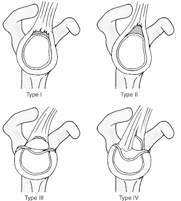
SLAP ( S uperior Labrum Anteriorly and Posteriorly  ) Lesions Compressive loading of the shoulder in the flexed abducted position (e.g. in a fall on the outstretched hand) can damage the superior labrum anteriorly and posteriorly (SLAP). The injury of the superior labrum begins posteriorly and extends anteriorly, stopping before or at the mid-glenoid notch and including the ‘anchor’ of the biceps tendon to the labrum. Four Main Types Are Described: Non-traumatic superior labral Degeneration , usually in older people and often asymptomatic; Avulsion of the superior part of the labrum – the commonest type A ‘ Bucket handle’ tear of the superior labrum; As for type 3 with an extension into the tendon  of long head of biceps. • Further subtypes that include associated lesions have also been described.  Clinical Features There is usually a history...

Popular posts from this blog

SOMI Brace

Skeletal Traction: Common sites

Pavlik Harness