Posts

Showing posts from March, 2017

Hip Joint: History Taking (Long Case)

My patient Mr. X, son of Mr. Y, 40 years old male, labourer by occupation, Resident of Z, presented with the CHIEF COMPLAINTS of Pain in right hip for 3 years Difficulty in walking /weight bearing for 3 years Limp for 3 years Deformity of right hip/shortening of right lower limb for 2 years Restriction of movements (stiffness) of right hip for 6 months HOPI (History of Presenting illness) - as stated by the patient Patient was apparently asymptomatic 3 years back when he developed PAIN over his right hip, which was insidious in onset, gradually progressive, continuous/intermittent, mild to moderate in intensity, stabbing/pricking/dull aching in nature, non-radiating, aggravated by wt bearing/movement/walking, relived by taking rest/walking and medications. There was no diurnal or seasonal variation. Pain was associated with DIFFICULTY IN WEIGHT BEARING . Initially patient was ab...

Skeletal Traction: Common sites

Image
1. Olecranon Site: 1.25 inches (3 cm) distal to the tip of olecranon just deep to the subcutaneous border of upper end of the ulna. Direction:  Medial to lateral at right angles to the longitudinal axis of ulna. Avoid: Ulnar nerve injury. 2. Second & Third Metacarpals Site:  About 1 inch (2-2.5 cm) Proximal to the distal end of Second metacarpal Direction:  The wire traverses 2nd and 3rd metacarpals transversely at right angles to the longitudinal axis of the radius. 3. Greater trochanter (upper end of femur ) Site: ...

Magnetic Resonance Imaging (MRI) in Orthopaedics

Image
Magnetic resonance imaging  produces cross-sectional images of any body part in any plane . It yields  superb soft-tissue contrast , allowing different soft tissues to be clearly distinguished, e.g. ligaments, tendons, muscle and hyaline cartilage. Another big advantage of MRI is that  it does not use  ionising  radiation. MRI Physics   The patient’s body is placed in a strong magnetic field (between 5 and 30,000 times the strength of the earth’s magnetic field). ⬇ The body’s protons have a positive charge and align themselves along this strong external magnetic field. ⬇ The protons are spinning and can be further excited by radio frequency pulses, rather like whipping a spinning top. ⬇ These spinning positive charges will not only induce a small magnetic field of their own, but will produce a signal as they relax (slow down) at different rates. ⬇ A proton density map is recorded from these signal...

SLAP Lesions

Image
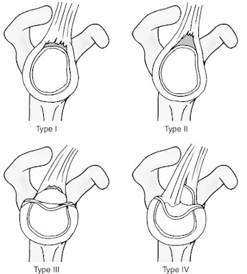
SLAP ( S uperior Labrum Anteriorly and Posteriorly  ) Lesions Compressive loading of the shoulder in the flexed abducted position (e.g. in a fall on the outstretched hand) can damage the superior labrum anteriorly and posteriorly (SLAP). The injury of the superior labrum begins posteriorly and extends anteriorly, stopping before or at the mid-glenoid notch and including the ‘anchor’ of the biceps tendon to the labrum. Four Main Types Are Described: Non-traumatic superior labral Degeneration , usually in older people and often asymptomatic; Avulsion of the superior part of the labrum – the commonest type A ‘ Bucket handle’ tear of the superior labrum; As for type 3 with an extension into the tendon  of long head of biceps. • Further subtypes that include associated lesions have also been described.  Clinical Features There is usually a history...

SOMI Brace

Image
Sternal-Occipital-Mandibular Immobiliser  (SOMI) Brace  Cervico-Thoracic Orthoses (CTOs) provide greater motion restriction from C5-C7 spine from the increased leverage on the person’s body. The upper cervical spine has less motion restriction. CTOs are used in minimally unstable fractures . All CTO’s tend to control flexion better than extension . Parts 1. The SOMI is a rigid, 3-poster CTO that has an padded anterior chest plate extending to the xiphoid process, as well as two padded shoulder extension which hook over tops of the shoulder. 2. From these shoulder extension two Straps which cross in the interscapular region, pass downward and around the chest wall to attach to the lower part of the chest plate. 3. There are three adjustable uprights , which pass upward from the chest plate, two to the padded occipital support and one to the mandibular support. 4. A removable chin piece attaches...

Skin Grafts

Image
Some wounds are not amenable to management by primary, delayed primary, or secondary closure. In such cases skin grafts can be used for secure wound coverage Definition -  A skin graft is a piece of skin taken from one part of the body , detached from its blood supply, and then transferred to another part of the body that allows its re-incorporation. Early adhesion of the graft is due to fibrin clot that forms within minutes after skin transfer The process of a graft being accepted at the recipient site is called ‘ Take ’. Steps of Graft uptake- Imbibition (24–48 h)— During this stage, the graft relies on nutrients reaching it by passive means  from the recipient bed . The graft increases in weight and volume.  Inosculation —Some vascularity is re-established by blood vessels in the recipient bed connecting to vessels in the graft dermis. Revascularization —after 4–7 days, new vessels form due to host endothelium ingrowth into t...

Popular posts from this blog

Hanging Cast

Skeletal Traction: Common sites

SOMI Brace