Pelvic Support Osteotomy


The pelvic support osteotomy is a useful surgical procedure for the salvage of damaged hips of patients in whom arthrodesis or hip arthroplasty are not appropriate.


The surgery is a double-level osteotomy of the femur:


(a) The more proximal valgus-extension osteotomy is performed with the femur in maximum adduction and at a level where the femoral shaft is seen to abut the pelvis.

(b) The second, more distal, osteotomy restores the orientation of the knee and ankle joint lines in the coronal plane and also provides a focus for femoral lengthening if warranted.


✫The proximal osteotomy lateralizes and distally displaces the greater trochanter and in doing so increases the action of the abductor muscles.

✫To this is added the elimination of any further adduction between femur and pelvis which then prevents pelvic drop during the single stance phase of gait.

✫ A successful pelvic support osteotomy
  1. Reduces limp through abolishing the Trendelenburg lurch,
  2. Equalises limb length and,
  3. Through the stability provided to the hemipelvis, facilitates a more energy-efficient gait.

Historical Aspect

The term ‘pelvic support’ is attributed to Lance who, in 1936, used it in reference to subtrochanteric osteotomies for the treatment of congenital dislocation of the hip.


✫ Variations of the procedure had been described in which it was believed that medial displacement of the anatomical axis of the femur in relation to the mechanical axis produced increased stability.
  • Milch contrasted the ideologies behind the variations and told that abutment of the upper end of the osteotomised shaft of femur against the pelvis was responsible.
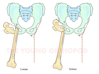
  • The techniques by Lorenz , Schanz and Ilizarov deserve special mention.

  • The subtrochanteric osteotomy designed by LORENZ was a valgus osteotomy coupled to a medial and proximal displacement of the shaft of femur
    • →  The almost vertical disposition of the femoral shaft ‘supported’ the pelvis from abutment.
    • →  However, the prominence of the displaced femoral shaft was noted to produce limitation of movement owing to the very same impingement against the lateral wall of the pelvis.
    • →  This improved when the prominence remodelled with time or was surgically removed

  • In contrast, the SCHANZ Osteotomy was performed by introducing a valgus, and sometimes extension, position to the distal femoral segment but without the proximal displacement of the Lorenz procedure.


→ Whilst this increased pelvic stability, it shared the same effect of abutment from the apex of angulation against the lateral wall of the pelvis, especially when the patient attempted to bring the widely abducted leg parallel to the opposite side.


  Both techniques therefore induced a limitation of movement from abutment.

MILCH highlighted this conundrum by introducing the concept of a POST-OSTEOTOMY ANGLE and its relation to pelvic inclination at the lateral wall of the ischium
   

  → When this angle, whether from Schanz or Lorenz type osteotomies, exceeded pelvic inclinationimpingement occurred when the patient attempted to bring the leg into parallel with the contralateral side


The loss of parallelism was in effect an ‘abduction contracture’ and meant some patients, when standing, had to compensate with eversion of the foot and with tilting of the pelvis


→ Whilst this had the desired effect of eliminating the Trendelenburg gait, excessive increase in the abduction angle (and consequently the post-osteotomy angulation) led to disability.

✫ Worse still, when the procedure was performed for bilateral cases this made compensation by pelvic tilting impossible.

 Milch recommended this post-osteotomy angle should lie between 210° and 240°

Ilizarov's distal osteotomy

The pelvic support osteotomy as described by ILIZAROV provided a solution through a second, more distal, osteotomy.

→ The significance of this additional osteotomy was to enable a proximal valgus osteotomy large enough to eradicate any degree of adduction in the hip (and thereby eliminate the Trendelenburg gait) but, through the distal varus osteotomy, achieve parallelism of both legs.


• If lengthening was performed through the distal osteotomy site, as was advocated by Ilizarov, parallelism of the both limbs with a level pelvis on standing was accomplished


Indications For Pelvic Support Osteotomy


Relative Contraindications

  1. Rapid remodelling at the proximal femoral osteotomy site should be anticipated if the procedure is performed in young children (under the age of 12 years), with the loss of pelvic support occurring as early as 12 months after the procedure.
  2. The procedure is also less suitable for older patients in whom THR are a better alternative.
  3. Further contraindications are chronic paralytic hip dislocations (from neuromuscular disorders, e.g. cerebral palsy, myelomeningocele, poliomyelitis) in non-ambulating patients.


Proximal Femoral Osteotomy:


a) Level Of Osteotomy
  • When the femoral shaft is fully adducted against the lateral wall of the pelvis, an AP X-ray of the pelvis gives a projection of abutment and this depicts the level of the proximal osteotomy.
b) Amount Of Valgus
  • The abduction angle should be either equal to the single stance pelvic drop angle or the measured range of adduction, plus an overcorrection factor of 15°–25°.
c) Amount Of Extension

✫ The second component of the proximal femoral osteotomy is extension to overcome the effects of a fixed flexion contracture of the hip.

  • A full correction of the fixed flexion deformity, without due consideration of the arc of hip flexion can be disadvantageous.

  • If the arc is small, some consideration to the loss of maximum hip flexion is needed
    → whilst patients may benefit from a better standing posture (having reduced or eliminated their lumbar lordosis), they may complain from being unable to fasten on their shoes.
  • Certainly a proportion of the fixed flexion contracture can be compensated for in the osteotomy and this usually amounts to 20°.

Distal Femoral Osteotomy:


a) Level Of Osteotomy
    • This second osteotomy is Ilizarov’s contribution to the pelvic support technique that addresses the excessive valgus of the proximal osteotomy and allows for derotation and lengthening as well

    ✫ Some advocate the level of the distal osteotomy be placed such that after varus correction, the centre of the knee joint is the same distance from the midline of the body as compared with the contralateral side.

b) Amount Of Varus
  • It was described that, in single stance, the ankle and knee joint inclinations in the coronal plane should be horizontal and parallel to the pelvis.
  1. →  Therefore this osteotomy serves to bring the inclination of the knee joint parallel with that of the horizontal line of the pelvis.
  2. →  However doing this will effectively remove any degree of overcorrection that is intentioned in the surgical planning.
  • To maintain some overcorrection, we suggest that the distal osteotomy is undercorrected (the knee joint line left in some valgus created by the proximal osteotomy)
  1. a convenient method is to leave the femoral shaft parallel to the vertical axis of the pelvis, thereby producing a valgus overcorrection of 9°–10°.
  • This explains the trigonometric method above which aims to bring the femoral shaft parallel to the vertical midline axis, thereby leaving a valgus inclination at the knee of 9° which is equivalent to an ‘abduction contracture’ of the same amount.
c) Amount Of Derotation

  • This can be performed at the distal osteotomy instead of the proximal. 
d) Amount Of Lengthening
  • The parallel beam scanogram provided an estimate of the length discrepancy between the limbs.
  • The most reliable measure of this difference is performed after the pelvic support osteotomy is carried out.
  • A new scanogram is needed and, together with a clinical estimate using blocks, the leg length discrepancy should be evaluated again.

✫  Lengthening is performed through the second osteotomy site in accordance with the principles laid down by Ilizarov.

✫  Over-lengthening is to be avoided as it is poorly tolerated in a hip that is already in full adduction. 

Comments

  1. The pelvic support osteotomy is a useful surgical procedure for the salvage of damaged hips of patients in whom arthrodesis or hip arthroplasty are not appropriate.
    Total Knee Replacement in Bangalore | Total Hip Replacement in Bangalore

    ReplyDelete

Post a Comment

Popular posts from this blog

Hanging Cast

SOMI Brace

Skeletal Traction: Common sites